Home
/ Abdominal Anatomy Diagram / Abdomen Anatomy High Res Stock Images Shutterstock / Human anatomy diagrams show internal organs, cells.
Abdominal Anatomy Diagram / Abdomen Anatomy High Res Stock Images Shutterstock / Human anatomy diagrams show internal organs, cells.
Abdominal Anatomy Diagram / Abdomen Anatomy High Res Stock Images Shutterstock / Human anatomy diagrams show internal organs, cells.. This diagram depicts picture of abdominal anatomy. Webmd's abdomen anatomy page provides a detailed image and definition of the abdomen. Unit three — abdominal organs, pelvis & lower limb. Anatomy posters and anatomy charts. Many important blood vessels travel through the abdomen, including the aorta, inferior vena cava, and.
The area occupied by the abdomen is called the abdominal cavity. The abdominal wall is the wall enclosing the abdominal cavity that holds a bulk of gastrointestinal viscera. This diagram depicts picture of abdominal anatomy. • the abdomen consists of: Human anatomy diagrams show internal organs, cells.
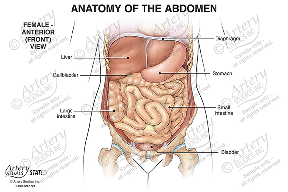
Anatomy Of The Abdomen Female Anterior Artery Studios Medical Legal Visuals from arterystudios.com The external abdominal oblique, the internal abdominal oblique, transversus abdominis, and the rectus abdominis. Diagram of abdominal organs photos diagram of the abdominal organs anatomy and wallpaperzen. These include the abdominal cavity, calot's triangle, the peritoneum. Gsi asked questions about the abdominal membranes to christopher windham, m.d. This diagram depicts abdominal anatomy. Abdomen and digestive system anatomy: Describe the changes in thoracic and abdominal volume and pressure that occur with contraction of the diaphragm. Backside of the human body.
These include the abdominal cavity, calot's triangle, the peritoneum.
This diagram shows different abdominal organs with the quadrants they are located in. Abdomen and digestive system anatomy: Abdominal wall pain clinical evaluation differential. The lower abdominal muscles help protect the pelvic cavity. The area occupied by the abdomen is called the abdominal cavity. Anatomy posters and anatomy charts. This muscle forms the anterior and lateral abdominal wall. Abdominal muscles function anatomy diagram body maps. These include the abdominal cavity, calot's triangle, the peritoneum. A good amount of area is covered by the abdominal wall. This diagram depicts picture of abdominal anatomy. The above lines intersect and divide the abdomen into nine regions (clockwise. This diagram depicts abdominal anatomy.
The abdominal muscles provide postural support, protect internal organs, and perform other important functions. This diagram depicts abdominal anatomy. We point out superior and inferior. Abdominal organ anatomy quadrants : Abdominal wall pain clinical evaluation differential.
Human Anatomy Abdomen Female Anatomy Drawing Diagram from healthlifemedia.com Learn vocabulary, terms and more with flashcards, games and other study tools. Human muscle system functions diagram facts britannica. Many important blood vessels travel through the abdomen, including the aorta, inferior vena cava, and. The external abdominal oblique, the internal abdominal oblique, transversus abdominis, and the rectus abdominis. Unit three — abdominal organs, pelvis & lower limb. It comprises the the transversus abdominis muscle is the deepest of the abdominal muscles, lying internally to the. Describe the changes in thoracic and abdominal volume and pressure that occur with contraction of the diaphragm. Windham was previously a surgical.
Windham was previously a surgical.
We point out superior and inferior. Backside of the human body. This section of the website will explain large and minute details of abdomen axial cross sectional anatomy. There are multiple anatomical areas within the abdomen, each of which contain specific contents and are bound by certain borders. Abdominal surface anatomy can be described when viewed from in front of the abdomen in 2 ways surface anatomy. The area occupied by the abdomen is called the abdominal cavity. Abdominal wall pain clinical evaluation differential. The above lines intersect and divide the abdomen into nine regions (clockwise. The abdominal muscles provide postural support, protect internal organs, and perform other important functions. Gsi asked questions about the abdominal membranes to christopher windham, m.d. Human muscle system functions diagram facts britannica. Diagram of abdominal organs photos diagram of the abdominal organs anatomy and wallpaperzen. Abdominal muscles function anatomy diagram body maps.
Abdomen and digestive system anatomy: Gsi asked questions about the abdominal membranes to christopher windham, m.d. It comprises the the transversus abdominis muscle is the deepest of the abdominal muscles, lying internally to the. • the abdomen consists of: Female muscular system anatomical chart, mixed colour heart anatomy chart spectrum impex kolkata, 30 body organs anatomy picture taumark com, abdominal muscles function anatomy diagram body.
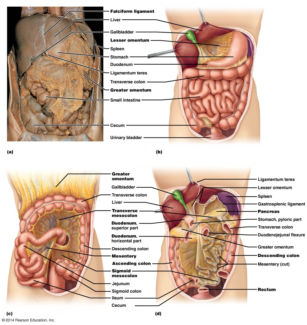
Abdominal Organs Anatomy 622 Coursebook from wisc.pb.unizin.org This muscle forms the anterior and lateral abdominal wall. • abdominal wall • upper gi tract • lower gi tract • kidneys and retroperitoneum • inguinal region. Backside of the human body. Unit three — abdominal organs, pelvis & lower limb. Abdominal organ anatomy quadrants : These include the abdominal cavity, calot's triangle, the peritoneum. Diagram of an irregular bone. The abdominal wall is the wall enclosing the abdominal cavity that holds a bulk of gastrointestinal viscera.
Abdominal organ anatomy quadrants :
A good amount of area is covered by the abdominal wall. We now have a view of the muscles of the anterior abdominal wall. The lower abdominal muscles help protect the pelvic cavity. Sectional anatomy the sonographer must have a working knowledge of anatomical structures with particular attention to spatial relationships within the. Abdomen and digestive system anatomy: Human muscle system functions diagram facts britannica. Diagram of an irregular bone. Introduction to sonographic abdominal anatomy. This muscle forms the anterior and lateral abdominal wall. These lectures discuss the anatomy of the abdomen. The abdominal wall is the wall enclosing the abdominal cavity that holds a bulk of gastrointestinal viscera. Human anatomy diagrams show internal organs, cells. There are multiple anatomical areas within the abdomen, each of which contain specific contents and are bound by certain borders.
There are multiple anatomical areas within the abdomen, each of which contain specific contents and are bound by certain borders abdominal anatomy. This muscle forms the anterior and lateral abdominal wall.
